

2 hours ago6 min read
2 days ago6 min read
4 days ago5 min read
Updated: Jan 5
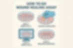
The wound healing assay, commonly known as the scratch assay, is a standard in vitro technique used to study collective cell migration and quantify the effects of various treatments or conditions on cell motility. This protocol provides a step-by-step guide for performing a reproducible wound healing assay using common laboratory equipment.
This method relies on creating a defined, cell-free gap ("wound" or "scratch") within a confluent monolayer of cultured cells. The rate at which cells migrate from the edges to close this gap is monitored over time, typically using time-lapse microscopy. Comparing the closure rates between different experimental conditions allows researchers to assess factors influencing cell migration.
Appropriate cell culture medium (e.g., DMEM, RPMI) supplemented with Fetal Bovine Serum (FBS) and antibiotics (e.g., Penicillin/Streptomycin) as required for the specific cell line.
Phosphate-Buffered Saline (PBS), sterile, Ca++/Mg++ free.
Trypsin-EDTA solution for cell detachment.
(Optional) Mitomycin C or other proliferation inhibitor.
(Optional) Cell staining reagents (e.g., Crystal Violet, Hoechst) if endpoint analysis is performed.
Experimental compounds (inhibitors, activators, drugs) dissolved in appropriate vehicle. Control medium containing only the vehicle.
Sterile tissue culture plates (e.g., 6-well, 12-well, 24-well, or 96-well plates are commonly used). Choose based on experimental needs and imaging capabilities.
Sterile pipette tips (P1000, P200, P20, P10 as needed). A P200 or P10 tip is often used for creating the scratch.
Serological pipettes.
Cell scraper or rubber policeman (if needed for harvesting).
Standard cell culture incubator (37°C, 5% CO2, humidified).
Biological safety cabinet (BSC).
Inverted microscope equipped with a camera (phase contrast optics recommended).
(Optional but recommended) Live-cell imaging system with environmental control (temperature, CO2, humidity) for time-lapse microscopy.
Centrifuge.
Hemocytometer or automated cell counter.
Water bath (for warming media).
Harvest logarithmically growing cells using standard trypsinization methods.
Count cells accurately using a hemocytometer or cell counter.
Seed cells into the chosen culture plate wells at a density determined (previously optimized) to achieve 95-100% confluence within 24-48 hours. Even cell distribution is crucial.
Example density: For a 24-well plate, ~0.1 x 10^6 cells per well might be appropriate, but this must be optimized per cell line.
Incubate cells under standard conditions (37°C, 5% CO2) until a confluent monolayer is formed.
If desired, serum-starve cells for a few hours (e.g., 2-4 hours) before scratching to synchronize cell cycles.
If inhibiting proliferation is necessary (to ensure gap closure is primarily due to migration), treat cells with Mitomycin C (e.g., 10 µg/mL for 1-2 hours) before creating the scratch, following established protocols for your cell line. Wash thoroughly after treatment.
Once cells are confluent (and optionally pre-treated), carefully remove the culture medium.
Using a sterile P200 or P10 pipette tip, create a straight scratch across the center of the monolayer. Apply firm, consistent pressure and speed.
Tip: Hold the pipette vertically. Aligning the tip with a marking on the plate bottom or using a ruler/guide can aid consistency. Multiple scratches per well can be made if needed, ensuring they are parallel.
Alternatively, use CytCut to create consistent scratches or commercially available wound healing assay inserts or stoppers which create a defined gap without physically scratching the surface.
Gently wash the wells 2-3 times with sterile PBS or serum-free medium to remove detached cells and debris created by the scratch. Perform washes carefully to avoid disturbing the monolayer edges. Aspirate the final wash.
Add fresh culture medium to each well. This medium should contain the experimental treatments (e.g., drug, inhibitor, growth factor) or vehicle control.
Often, medium with reduced serum (e.g., 0.5-2% FBS) is used during the migration phase to minimize proliferation effects if Mitomycin C was not used.
Immediately place the plate on the microscope stage (ideally within a live-cell incubator).
Capture the first image (T=0) of the scratch in predefined locations for each well. Use phase-contrast microscopy to visualize the cell-free gap clearly. Ensure consistent focus and lighting.
Continue capturing images at regular intervals (e.g., every 2, 4, 6, or 12 hours) for the duration of the experiment (typically 12-72 hours, depending on cell migration speed).
Marking reference points on the plate bottom outside the field of view can help ensure the same region is imaged each time.
Once time-lapse images have been acquired, quantitative analysis is performed to compare cell migration rates between experimental conditions. While traditional methods often focus on measuring the change in the area or width of the cell-free gap, an alternative approach using Relative Wound Density (RWD) offers advantages, particularly in accounting for cell proliferation and variations in cell density (as described by Topman et al., PLOS ONE, 2020).
The RWD method quantifies the extent to which the initial wound area has been repopulated by cells, relative to the density of the surrounding cell layer at the same time point.
Using image analysis software (e.g., ImageJ/Fiji, CellProfiler, or other suitable software), carefully delineate the boundary of the cell-free area in the image taken immediately after scratching (T=0). This defined area serves as the reference wound ROI for all subsequent time points for that specific well/field of view.
For each captured image at time t (including T=0 and subsequent time points), use the software's segmentation tools to distinguish between cell-covered areas and background (empty space). This might involve thresholding based on pixel intensity (common in phase contrast) or identifying stained nuclei if fluorescence imaging is used.
For each time point t:
Wound Density (Density_{wound}(t)): Measure the cell density specifically inside the initially defined wound ROI. This density can be expressed as the percentage of the ROI area covered by cells, or as the number of cells per unit area if individual cells/nuclei are counted.
Outside Density (Density_{outside}(t)): Measure the cell density in the region outside the initial wound ROI (i.e., the surrounding monolayer). This serves as an internal reference for the maximum achievable cell density at that time point, implicitly accounting for proliferation or density changes in the monolayer itself.
For each time point t, calculate the RWD using the following formula: RWD(t) = Density_{wound}(t) / Density_{outside}(t)
Note: At T=0, Density_{wound}(0) should ideally be close to 0, making RWD(0) near 0. As cells migrate into the wound, Density_{wound}(t) increases, and thus RWD(t) increases.
The RWD value ranges from approximately 0 (no closure) to 1 (complete closure, where the density inside the original wound matches the density outside).
Plot RWD(t) versus time for each experimental condition.
Compare the RWD curves: A faster increase in RWD indicates a faster rate of wound closure/cell migration relative to the surrounding cell density.
Use appropriate statistical analysis (e.g., comparing RWD values at specific time points or analyzing the slopes of the RWD curves) to determine significant differences between treatments.
This analysis requires image processing software capable of defining ROIs, performing robust cell segmentation (potentially requiring optimization of thresholding or learning-based algorithms), and measuring area coverage or object counts within specified regions. ImageJ/Fiji with appropriate plugins or custom macros is a common open-source option.
An alternative method to get the gap area of all your images without any complex analysis and in matters of seconds is Sophie Scratch analyzer. Just upload your t0 an secondary time images and get an excel output with all gap area percentages!
By using the RWD metric, the analysis normalizes the wound repopulation against the state of the surrounding monolayer, providing a potentially more robust measure of cell migration dynamics compared to simple area closure, especially in experiments where cell proliferation significantly contributes to changes in cell density over time.
Cell Confluence: Ensure consistent, high confluence before scratching. Over- or under-confluence affects migration.
Scratch Consistency: Practice making uniform scratches. Variations in width greatly impact results. Consider using tools like CytCut 3.0 for better constancy.
Control Proliferation: If proliferation is a concern, use Mitomycin C pre-treatment or maintain cells in low-serum medium during migration. Run controls to assess proliferation independently if needed.
Imaging: Maintain consistent focus and illumination. Choose time intervals appropriate for the cell line's migration speed.
Edge Effects: Avoid analyzing areas too close to the well edges if migration appears different there.
Analysis: Using a standardized tool like Soφ scratch analyzer, where all parameters are consistent across all images, would result in faster analysis and higher quality data.
This detailed protocol outlines the steps for conducting a wound healing (scratch) assay, a fundamental method for investigating collective cell migration. Careful optimization, consistent execution, and appropriate analysis are key to obtaining reliable and reproducible data on cell motility in vitro.
References
Abcam. Wound Healing Assay Protocol. https://www.abcam.com/en-us/technical-resources/protocols/wound-healing-assay (Accessed: May 4, 2025)
Jonkman, J. E., et al. (2014). An introduction to the wound healing assay using live-cell microscopy. Cell Adhesion & Migration, 8(5), 440-451.
https://journals.plos.org/plosone/article?id=10.1371/journal.pone.0232565